
-
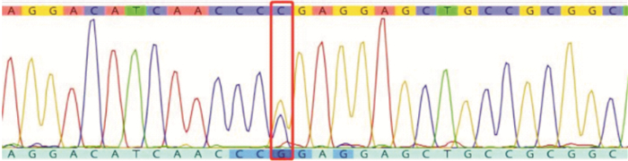
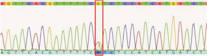
摘要: 目的 分析P1(-)表型个体产生抗P1抗体的血型、抗体鉴定的血清学检测结果、分子机制以及交叉配血策略,并回顾对比分析既往2例血清学检测方法和结果,对优化检测方法提高抗P1抗体检出率进行探讨。方法 采用盐水法、聚凝胺和微柱凝胶法进行血型鉴定、抗体筛选和交叉配血,采用PCR扩增α-1,4-半乳糖基转移酶基因(α-1,4-galactosyltransferase,A4GALT)和β-1,3-N-乙酰半乳糖氨基转移酶基因(β-13-N-acetylgalactosaminyltransferase,B3GALNT1)外显子编码区,分析DNA序列。结果 患者1经过盐水法、微柱凝胶法、抗人球蛋白法血清学分析鉴定为IgM抗P1抗体,经过PCR核酸检测确定为患者表型为P1(-);P型,验证与血清学的检测结果一致。患者2仅通过血清学检测鉴定为抗P1抗体。结论 抗P1抗体多为IgM型抗体,在进行抗体检测时需考虑到检测方法的适用范围和局限性,选择合适的方法,并进行多种方法联合检测相互验证的方式,以提高抗体的检出率。核酸检测基因分型技术的应用可以从分子生物学角度提供重要佐证,有助于提高抗体鉴定的准确率。Abstract: Objective To review the blood type of anti-P1 antibody produced by individuals with P1(-) phenotype, the serological test results of antibody identification, molecular mechanism and cross-matching strategy, compare the previous two serological test methods and results, and discuss the optimization of test methods to improve the detection rate of anti-P1 antibody.Methods Blood group identification, antibody screening and cross matching were performed by normal saline method, polybrene and microcolumn gel method, and PCR amplification was used α-1, 4-galactosyltransferase gene(A4GALT) and β-1, 3-N-acetylgalactosamine aminotransferase gene(B3GALNT1)exon coding region, and DNA sequence was analyzed.Results Patient 1 was identified as IgM anti-P1 antibody by normal saline method, microcolumn gel method and antiglobulin method, and the phenotype of patient 1 was P1(-) by PCR nucleic acid detection. P type, was verified to be consistent with the results of serological detection. Patient 2 was identified with anti-P1 antibodies only by serologic testing.Conclusion Most of the anti-P1 antibodies were IgM. When performing antibody detection, it is necessary to consider the scope and limitations of the detection methods, select the appropriate method, and conduct a variety of combined detection methods to verify each other, so as to improve the detection rate of antibody. The application of nucleic acid detection genotyping technology can provide important corroboration from a molecular biology perspective and help to improve the accuracy rate of antibody identification.
-
Key words:
- anti-P1 antibody /
- antibody identification /
- genotyping /
- comparison of methodology
-

-
表 1 2例ABO、RHD血型鉴定结果
编号 伯乐全自动血型分析仪IH-1000 盐水试管法 盐水试管法P1抗原鉴定 最终结果 抗-A 抗-B 抗-D Ac Bc ctrl 抗-A 抗-B 抗-D Ac Bc Oc Ac Bc Oc 患者 患者1 4+ 0 4+ 1+ 4+ 0 4+ 0 4+ 1+ 4+ 1+ P1 / / 0 A+ 患者2 0 4+ 4+ 4+ ± 0 0 4+ 4+ 4+ 1+ 1+ / P1 / 0 B+  下载: 导出CSV
表 2 患者血清与抗体筛选细胞反应结果
编号/试剂批号 检测方法 与筛选细胞反应格局 Sc 鉴定结果 Ⅰ Ⅱ Ⅲ 患者1/20220802 微柱凝胶 ± 3+ 0 0 抗M、S、P1 盐水法 3+ 4+ 0 0 抗M、S、P1 2-Me处理后 0 0 0 0 患者2/20200702 微柱凝胶 0 0 0 0 未检出 盐水法 0 0 + 0 抗Fyb、Leb、P1
下载: 导出CSV
表 3 患者血清与谱细胞反应鉴定结果
编号/试剂批号 检测方法 与谱细胞反应格局 Sc 鉴定结果 1 2 3 4 5 6 7 8 9 10 11 12 13 14 15 16 患者1/20220905 微柱凝胶 0 0 0 0 0 2+ 2+ 0 2+ 0 2+ 3+ 2+ 0 0 0 0 未确定 盐水法 2+ 2+ 0 2+ 2+ 2+ 2+ 0 2+ 0 2+ 2+ 2+ 2+ 0 2+ 0 抗P1 抗人球蛋白法 1+ 1+ 0 1+ 1+ 2+ 2+ 0 1+ 0 1+ 3+ 2+ 1+ 0 2+ 0 抗P1 患者2/20200611 微柱凝胶 / / / / / / / / / / 0 未检出 盐水法 ± 0 ± 0 2+ + ± + 0 ± 0 抗P1
下载: 导出CSV
-
[1] 龚道元. 临床输血检验技术(第2版/创新教材)[M]. 北京: 人民卫生出版社, 2021: 28-29.
[2] Mark KF. 美国血库协会技术手册[M]. 北京: 人民卫生出版社, 2020: 196-197.
[3] 徐路琼, 彭靖琪, 窦红勇, 等. p表型孕妇的妊娠管理[J]. 临床血液学杂志, 2021, 34(10): 732-736. https://lcxy.whuhzzs.com/article/doi/10.13201/j.issn.1004-2806.2021.10.012
[4] 屠佳燕, 周建华, 吴瑾惠, 等. p表型个体的血清学特点和分子机制研究1例[J]. 中华医学遗传学杂志, 2023, 40(3): 291-294.
[5] 魏玲, 姬艳丽, 骆宏, 等. 一例罕见P表型个体家系研究[J]. 中华医学遗传学杂志, 2012, 29(6): 701-704. doi: 10.3760/cma.j.issn.1003-9406.2012.06.017
[6] 许露萍, 尹俊皓, 王若涵. 罕见抗-Tj. (a)合并自身抗体致疑难配血分析[J]. 临床输血与检验, 2021, 23(6): 800-802. doi: 10.3969/j.issn.1671-2587.2021.06.028
[7] 刘世佳, 张勇萍, 杨世明, 等. IgM抗Le~a、抗P_1抗体联合IgG抗E抗体的血清学检测分析[J]. 细胞与分子免疫学杂志, 2021, 10: 932-935.
[8] 李丹, 杨琳, 杨世明, 等. 抗P_1抗体及其联合抗M、抗E抗体的鉴定分析[J]. 细胞与分子免疫学杂志, 2023, 9: 841-845. https://www.cnki.com.cn/Article/CJFDTOTAL-XBFM202309011.htm
[9] 石翠英, 刘敬闪, 王毅, 等. 天然抗-P1引起血型定型困难1例[J]. 中国输血杂志, 2000, 13(2): 132. https://www.cnki.com.cn/Article/CJFDTOTAL-BLOO200002047.htm
[10] 陈凯, 庄文华, 陈平, 等. 献血者抗-P1抗体的鉴定和处置策略探讨[J]. 医学检验与临床, 2022, 33(8): 66-67, 72.
[11] 赵梓昕, 许先国, 洪小珍, 等. 血清学和分子生物学鉴定罕见p表型——附1例报告[J]. 中国输血杂志, 2019, 32(11): 1116-1120.
[12] 张晨光, 张婧婧, 庞桂芝, 等. 血型不规则抗体检测的方法学评价[J]. 检验医学, 2010, 25(12): 929-933.
[13] Loua A, Nikiema JB, Sougou A, et al. Transfusion in the WHO African region[J]. Transfus Clin Biol, 2019, 26(3): 155-159.
[14] 王文婷, 顾顺利, 陈要臻, 等. 微柱凝胶卡交叉配血法导致血型抗体漏检的探讨[J]. 中国输血杂志, 2019, 32(7): 691-693. https://www.cnki.com.cn/Article/CJFDTOTAL-BLOO201907029.htm
[15] Smith D, Aye T, Er LS, et al. Acute hemolytic transfusion reaction due to anti-P1: a case report and review of institutional experience[J]. Transfus Med Hemother, 2019, 46(5): 380-383.
[16] Arndt PA, Garratty G, Marfoe RA, et al. An acute hemolytic transfusion reaction caused by an anti-P1 that reacted at 37 degrees C[J]. Transfusion, 1998, 38(4): 373-377.
[17] Bezirgiannidou Z, Christoforidou A, Kontekaki E, et al. Hyperhemolytic syndrome complicating a delayed hemolytic transfusion reaction due to anti-P1 alloimmunization, in a pregnant woman with HbO-Arab/β-thalassemia[J]. Mediterr J Hematol Infect Dis, 2016, 8(1): e2016053. http://europepmc.org/articles/PMC5111518?pdf=render
[18] Thakral B, Bhattacharya P, Agnihotri N, et al. Acute hemolytic transfusion reaction by anti-P1 antibody in pregnancy[J]. Am J Hematol, 2005, 78(2): 163-164.
-
图(2)
表(3)
计量
- 文章访问数: 518
- PDF下载数: 161
- 施引文献: 0