
Predictive value of thromboelastography combined with conventional coagulation indexes in risk of hypercoagulation in lung cancer patients
-
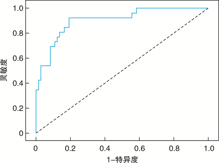
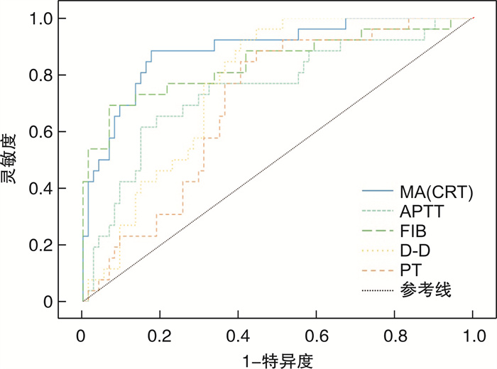
摘要: 目的 探讨血栓弹力图(TEG)联合常规凝血指标对肺癌患者凝血功能的检测与高凝状态的预测作用。方法 收集医院2021年1月1日—2023年3月31日收治的100例肺癌患者的TEG、常规凝血相关指标,并根据患者的凝血状态及临床表现分为高凝组(n=26)和非高凝组(n=74)。同时,选取40例健康者作为对照组,检测TEG指标:凝血反应时间(R值)、凝血形成速率(Angle)、凝血最大强度(MA)和常规凝血指标:凝血酶原时间(PT)、活化部分凝血活酶时间(APTT)、凝血酶时间(TT)、D-二聚体(D-D)、纤维蛋白原(FIB)。记录三组数据,分析不同指标之间的差异与相关性。结果 肺癌患者与健康者比较,其Angle、MA、APTT、FIB、D-D增大,而R值降低。高凝组、非高凝组与健康组三组均数比较,APTT、FIB、Angle、MA之间的差异有统计学意义(P < 0.05)。TEG与常规凝血五项指标之间的相关性在高凝组中发现,Angle与FIB呈正相关(P < 0.05),MA与FIB呈正相关(P < 0.05);R值与PT呈正相关(P < 0.05),与FIB呈负相关(P < 0.05)。通过受试者工作特征(ROC)曲线分析,联合TEG与常规凝血评估肺癌患者凝血状态的ROC曲线下面积为0.905,灵敏度为0.932,特异度为0.811,对患者高凝状态的评估效能最佳。结论 相较于传统的常规凝血指标,TEG能够更快、更准确地发现肺癌患者的凝血异常。在高凝状态下,常规凝血指标与TEG参数之间具有相关性。联合TEG与常规凝血指标共同评估,有助于发现肺癌患者的凝血异常,预测高凝风险,指导临床用药预防血栓的形成。Abstract: Objective To investigate the effect of thromboelastography(TEG) combined with conventional coagulation on the detection of coagulation function and the prediction of hypercoagulability in lung cancer patients.Methods A total of 100 lung cancer patients in our hospital from January 1, 2021 to March 31, 2023 were collected from TEG and routine coagulation-related indicators, and were divided into hypercoagulable group(n=26) and non-hypercoagulation group(n=74) according to their coagulation status and clinical manifestations. At the same time, 40 healthy individuals were selected as the control group to test TEG indicators: coagulation reaction time(R value), coagulation formation rate(Angle angle), maximum coagulation strength(MA), and conventional coagulation indicators: prothrombin time(PT), activated partial thromboplastin time(APTT), thrombin time(TT), D-dimer(D-D) and fibrinogen(FIB).Results Compared with healthy patients, the Angle, MA, APTT, FIB, and D-D values of lung cancer patients increased, while the R value decreased. There were statistically significant differences in APTT, FIB, Angle and MA between the hypercoagulable group, non-hypercoagulable group and healthy group(P < 0.05). The correlation between TEG and the five conventional coagulation indexes was found in the hypercoagulable group, and the Angle angle was positively correlated with FIB(P < 0.05), and the MA value was positively correlated with FIB(P < 0.05). The R value was positively correlated with PT(P < 0.05) and negatively correlated with FIB(P < 0.05). Analysis of receiver operating characteristic(ROC) curve by subject. The ROC area under the curve of lung cancer patients evaluated by combining TEG and conventional coagulation was 0.905, with a sensitivity of 0.932 and a specificity of 0.811. This was the best way to assess the patient's hypercoagulability.Conclusion Compared with the traditional four coagulation indexes, TEG can detect coagulation abnormalities in lung cancer patients faster and more accurately. In the hypercoagulable state, there was a correlation between conventional coagulation parameters and TEG parameters. Combined TEG and conventional coagulation indexes can help to detect coagulation abnormalities in lung cancer patients, predict the risk of hypercoagulation, and guide clinical medication to prevent the formation of thrombosis.
-
Key words:
- thromboelastography /
- routine coagulation index /
- lung cancer /
- hypercoagulable state
-

-
表 1 TEG检测指标结果
X±S 组别 Angle/° MA/mm R/s 高凝组 82.73±1.851)2) 76.57±5.371)2) 34.89±5.56 非高凝组 78.63±3.331) 64.89±6.661) 35.60±6.42 健康组 75.14±3.01 60.06±3.75 35.73±5.32 正常范围 64~80 52~71 22~44 与健康组比较,1)P < 0.05;与非高凝组比较,2)P < 0.05。  下载: 导出CSV
表 2 常规凝血五项检测指标结果
X±S 组别 APTT/s FIB/(g/L) PT/s TT/s D-D/(ng/mL) 高凝组 35.81±6.261)2) 6.01±1.871)2) 12.36±0.641) 16.34±1.442) 1 235.88±736.381)2) 非高凝组 30.65±5.26 3.73±1.011) 12.38±4.94 16.89±1.10 825.05±753.881) 健康组 30.98±3.57 2.82±0.71 11.84±0.81 16.67±0.71 331.73±66.88 正常范围 24~39 2~4 10~14 14~21 0~500 与健康组比较,1)P < 0.05;与非高凝组比较,2)P < 0.05。
下载: 导出CSV
表 3 高凝组TEG参数与常规凝血检测结果相关性
指标 Angle MA(CRT) R(CRT) r P r P r P APTT 0.327 0.103 0.570 0.136 0.051 0.041 FIB 0.118 0.039 0.084 0.047 -0.059 0.019 PT -0.026 0.616 -0.139 0.133 0.337 0.009 TT 0.028 0.890 0.172 0.402 0.098 0.634 D-D -0.292 0.147 -0.306 0.129 -0.005 0.981
下载: 导出CSV
-
[1] Wu FY, Wang L, Zhou CC. Lung cancer in China: current and prospect[J]. Curr Opin Oncol, 2021, 33(1): 40-46. doi: 10.1097/CCO.0000000000000703
[2] 张维, 邱惠, 丁贤彬, 等. 重庆市2014年恶性肿瘤流行病学数据分析及防治对策研究[J]. 重庆医学, 2017, 46(11): 1511-1512, 1515. https://www.cnki.com.cn/Article/CJFDTOTAL-CQYX201711028.htm
[3] Bayleyegn B, Adane T, Getawa S, et al. Coagulation parameters in lung cancer patients: a systematic review and meta-analysis[J]. J Clin Lab Anal, 2022, 36(7): e24550. doi: 10.1002/jcla.24550
[4] 赵乐乐, 曹晓红. 肿瘤患者的静脉血栓风险模型研究进展[J]. 临床肺科杂志, 2022, 27(10): 1588-1592. https://www.cnki.com.cn/Article/CJFDTOTAL-LCFK202210028.htm
[5] 马旭, 韩森, 聂鋆, 等. 肺癌合并静脉血栓栓塞症患者的诊疗特点[J]. 肿瘤防治研究, 2020, 47(5): 335-339. https://www.cnki.com.cn/Article/CJFDTOTAL-ZLFY202005004.htm
[6] 谢华琴, 胡芬, 付雪娇. 血栓弹力图在预防肺癌患者PICC相关性静脉血栓中的应用[J]. 现代预防医学, 2019, 46(24): 4523-4526. https://www.cnki.com.cn/Article/CJFDTOTAL-XDYF201924029.htm
[7] 朱丽华, 周蕾, 罗斌, 等. 284例非小细胞肺癌患者循环肿瘤细胞与临床特征及凝血功能的相关性[J]. 肿瘤预防与治疗, 2020, 33(9): 746-752.
[8] 曾覃平, 杜秀娟, 彭碧, 等. 血栓弹力图与常规凝血功能检测在肺癌患者凝血功能中的应用评价[J]. 检验医学与临床, 2019, 16(4): 514-516. https://www.cnki.com.cn/Article/CJFDTOTAL-JYYL201904024.htm
[9] 施珊娜, 刘晓静, 许萍, 等. 血栓弹力图联合分子标志物对肺癌患者凝血功能的评估价值及其与预后的关系[J]. 中国当代医药, 2023, 30(23): 7-11. https://www.cnki.com.cn/Article/CJFDTOTAL-ZGUD202323033.htm
[10] 杨言, 夏萍, 毛玉琴, 等. Caprini评分、D-二聚体联合血栓弹力图对中晚期肺癌患者静脉血栓栓塞症发生的预测价值分析[J]. 肿瘤综合治疗电子杂志, 2023, 9(3): 98-103. https://www.cnki.com.cn/Article/CJFDTOTAL-ZLZD202303016.htm
[11] 李廷廷, 战翠萍, 崔久嵬. 癌症相关性血栓形成的病理机制进展[J]. 中国实验诊断学, 2019, 23(5): 932-935. https://www.cnki.com.cn/Article/CJFDTOTAL-ZSZD201905055.htm
[12] Tang N, Jin X, Sun ZY, et al. Effects of hemolysis and lipemia interference on Kaolin-activated thromboelastography, and comparison with conventional coagulation tests[J]. Scand J Clin Lab Invest, 2017, 77(2): 98-103. doi: 10.1080/00365513.2016.1271906
[13] 张宇, 阴鑫哲, 李林军, 等. 脊柱手术后预防下肢深静脉血栓形成抗凝方案研究[J]. 血栓与止血学, 2022, 28(3): 464-465. https://www.cnki.com.cn/Article/CJFDTOTAL-XSZX202203046.htm
[14] 郭莹莹, 赵震, 孙长杰, 等. 血栓弹力图在评估肝硬化患者凝血功能中的应用[J]. 中国实验诊断学, 2022, 26(12): 1804-1806. https://www.cnki.com.cn/Article/CJFDTOTAL-ZSZD202212017.htm
[15] Holcomb JB, Minei KM, Scerbo ML, et al. Admission rapid thrombelastography can replace conventional coagulation tests in the emergency department: experience with 1974 consecutive trauma patients[J]. Ann Surg, 2012, 256(3): 476-486. doi: 10.1097/SLA.0b013e3182658180
[16] 饶欣, 闫寒. 血栓弹力图参数预测急诊重症颅脑损伤患者并消化道出血的研究[J]. 临床急诊杂志, 2023, 24(7): 335-339. https://www.cnki.com.cn/Article/CJFDTOTAL-ZZLC202307001.htm
[17] 曹梦霞, 李凤娟, 任璐, 等. 基于血栓弹力图的凝块溶解时间与ACS合并高脂血症患者再发心肌梗死的相关性研究[J]. 临床心血管病杂志, 2022, 38(5): 378-383. https://www.cnki.com.cn/Article/CJFDTOTAL-LCXB202205009.htm
[18] 禹梅, 李娜, 寇长元, 等. 血栓弹力图评估胃癌患者围手术期凝血状态的应用研究[J]. 现代生物医学进展, 2017, 17(14): 2721-2724. https://www.cnki.com.cn/Article/CJFDTOTAL-SWCX201714030.htm
[19] 荆晶, 王文婷, 常艳, 等. 临床凝血功能异常患者血栓弹力图与常规凝血检测的比较及相关性分析[J]. 中国实验血液学杂志, 2020, 28(2): 629-635. https://www.cnki.com.cn/Article/CJFDTOTAL-XYSY202002050.htm
[20] 丁浩然, 胡艳, 李纲, 等. 血栓弹力图和常规凝血检验在老年慢性肾脏病检查中的应用[J]. 老年医学与保健, 2023, 29(6): 1294-1297. https://www.cnki.com.cn/Article/CJFDTOTAL-LYBJ202306038.htm
[21] Cotton BA, Minei KM, Radwan ZA, et al. Admission rapid thrombelastography predicts development of pulmonary embolism in trauma patients[J]. J Trauma Acute Care Surg, 2012, 72(6): 1470-1475;discussion 1475-1477. doi: 10.1097/TA.0b013e31824d56ad
[22] 蔡丹, 黄向红, 张远芳, 等. 血栓弹力图与常规凝血检测评估早期复发性流产患者凝血功能及指导抗凝治疗的预后比较[J]. 生殖医学杂志, 2023, 32(1): 55-60. https://www.cnki.com.cn/Article/CJFDTOTAL-SZYX202301009.htm
[23] 贺扬欣, 张萌, 崔颖, 等. 血栓弹力图与传统凝血指标预测脑卒中合并静脉血栓栓塞症的临床价值[J]. 西部医学, 2024, 36(1): 91-96, 102. https://www.cnki.com.cn/Article/CJFDTOTAL-XIBU202401017.htm
[24] 黄莹, 许燕燕, 王贤群, 等. 评估老年骨折患者凝血状态常用方法的对比研究[J]. 临床血液学杂志, 2023, 36(2): 113-116. doi: 10.13201/j.issn.1004-2806.2023.02.008
[25] 郝鹏飞, 高智, 党培业, 等. 抗凝时间对老年髋部骨折患者围术期深静脉血栓发生率的影响[J]. 血栓与止血学, 2022, 28(2): 290-291. https://www.cnki.com.cn/Article/CJFDTOTAL-XSZX202202050.htm
-
图(2)
表(3)
计量
- 文章访问数: 659
- PDF下载数: 776
- 施引文献: 0